Wrist | Carpal fractures | Hamate fracture
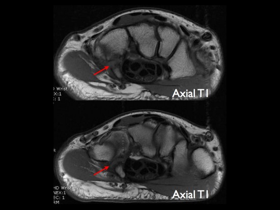
Fracture of the hamate are uncommon (2% of the wristt fractures) and occur most often at the base of the hamulus (or hook). Axial T1-weighted images:
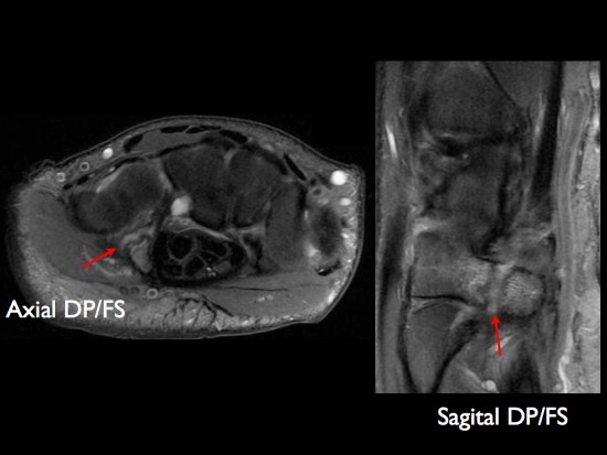
Sagittal and axial PD-weighted images with fat-saturation show hamuls fracture with bone marrow edema of the most anterior fragment.
CT (case 2):
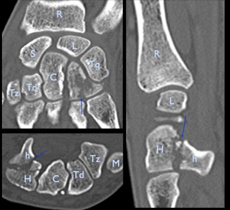
R: Radius - S: Scaphoid - L: Lunatum - Tq: Triquetrum - C: Capitatum - Tz: Trapezium - Td: Trapezoid
H: Hamatum - h: hamulus - M: metacarpal bone of the thumb
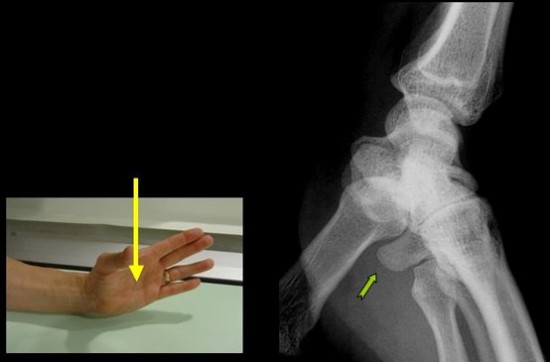
X-rays are often normal and oblique views are required to better assess the hamulus.