
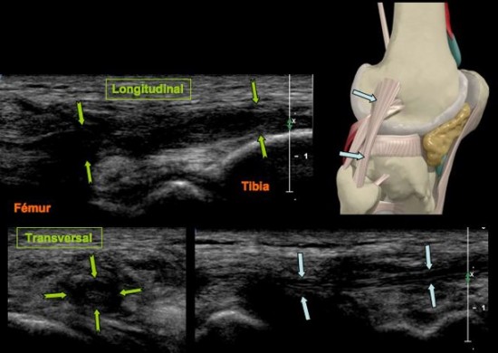
Genou | Ligaments collatéraux
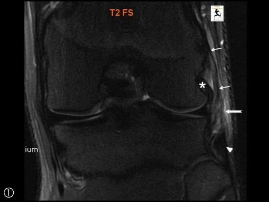
Mouvement forcé en valgus lors d’un raffût au rugby.
Désinsertion du faisceau profond, ménisco-fémoral, (![]() ) du Ligament Collatéral Médial (LCM) avec hyperhémie locale (
) du Ligament Collatéral Médial (LCM) avec hyperhémie locale (![]() ) . Le faisceau superficiel est continu (
) . Le faisceau superficiel est continu (![]() ), de même que l’insertion tibiale du ligament (
), de même que l’insertion tibiale du ligament (![]() ).
).
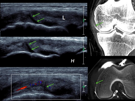
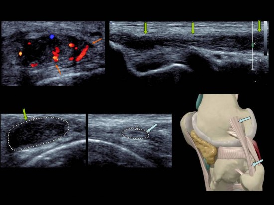
Echographie: image linéaire hypoéchogène non transfixiante (flèche verte) au sein du ligament collatéral médial (L) qui est épaissi. Il existe une hyperhémie au doppler couleur. Cette anomalie est mieux visible en mode harmonique (H).
Arthroscanner (réalisé avant échographie): image d'addition linéaire de produite de contraste à point de départ intra-articulaire et située au sein du ligament collatéral médial. Pas d'anomalie méniscale ou chondrale.
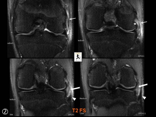
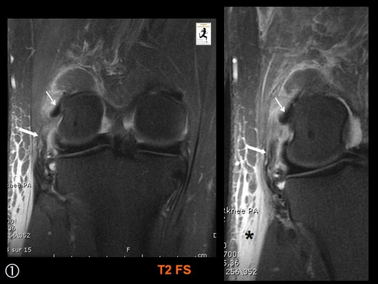
Entorse à Ski.
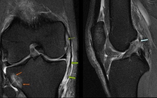
Rupture avec désinsertion distale ( ![]() ) du Ligament Collatéral Médial du Genou. Au niveau de son insertion proximale il existe également une désinsertion de son faisceau profond ménisco-fémoral (
) du Ligament Collatéral Médial du Genou. Au niveau de son insertion proximale il existe également une désinsertion de son faisceau profond ménisco-fémoral ( ![]() ). Rupture haute du Ligament Croisé Antérieur (
). Rupture haute du Ligament Croisé Antérieur ( ![]() ) et contusion osseuse de l'articulation Tibio Fibulaire Proximale (
) et contusion osseuse de l'articulation Tibio Fibulaire Proximale ( ![]() ).
).
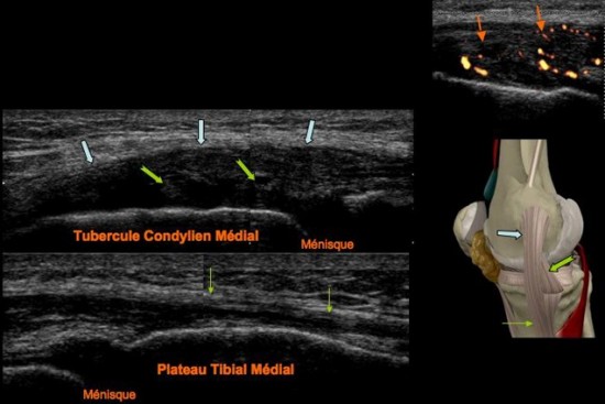
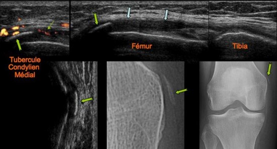
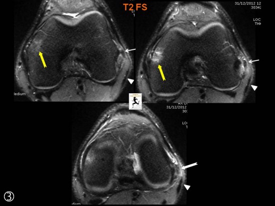
Aspect échographique normal du faisceau ménisco-fémoral ![]() du ligament collatéral médial,
du ligament collatéral médial,
faisceau superficiel normal ![]() et ménisque interne (MI).
et ménisque interne (MI).
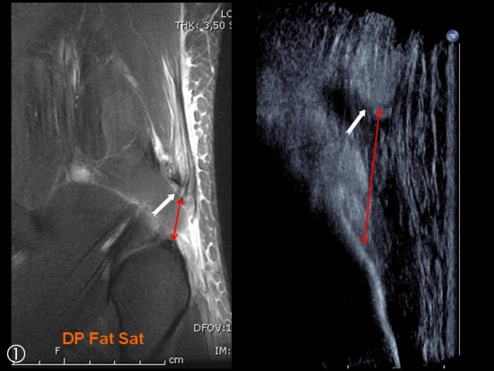
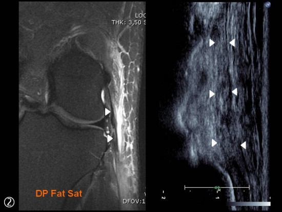
Traumatisme récent avec douleur du compartiment fémoro-tibial médial chez un rugbymen.
Epaississement proximal du ligament colatéral médial (![]() ) avec présence d’une ossification en regard du tubercule condylien médial retrouvé sur le bilan radiographique (
) avec présence d’une ossification en regard du tubercule condylien médial retrouvé sur le bilan radiographique (![]() ). La zone de cicatrisation est le siège d’une discrète hyperhémie au doppler couleur (
). La zone de cicatrisation est le siège d’une discrète hyperhémie au doppler couleur (![]() ).
).
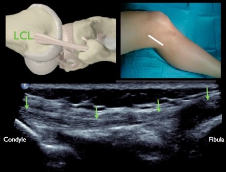
L’examen s’effectue en discrète flexion ce qui permet de «positionner» le ligament dans l’axe de la fibula.
Compte-tenu de la complexité de l’insertion distale du biceps qui «cravate» le ligament, la visibilité de l’insertion distale est souvent difficile.
Douleur du compartiment latéral après traumatisme au judo.
Distension ecchymotique (![]() ) isolée et sans rupture du LCL. Aspect normal du ligament opposé (
) isolée et sans rupture du LCL. Aspect normal du ligament opposé (![]() ).
).
Mouvement forcé au décours d'un match de rugby. Douleur du compartiment latéral.
Hypertrophie avec perte de l'architecture fibrillaire du Ligament Collatéral Latéral (![]() ) qui reste cependant continu.Importante hyperhémie au doppler couleur (
) qui reste cependant continu.Importante hyperhémie au doppler couleur ( ![]() ). Aspect normal du LCL opposé (
). Aspect normal du LCL opposé ( ![]() ). Le reste du compartiment latéral était sans anomalie. Pas de laxité a l'examen clinique.
). Le reste du compartiment latéral était sans anomalie. Pas de laxité a l'examen clinique.
Douleur postéro latérale du genou survenue après un tacle au football.
Rupture distale isolée (flèche blanche) du Ligament Collatéral Latéral (petites flèches blanches).
Les tendons Poplité (*) et du Biceps Fémoral (têtes de flèche blanches) étaient normaux.
La seule lésion associée était une petite plage de contusion osseuse du condyle médial (flèche jaune).
Traumatisme en hyperextension au rugby.
Désinsertion avec rétraction minime (flèches blanches) du Biceps Fémoral associée à une lésion intersticielle distale du Ligament Colatéral Latéral (têtes de fleches). Les deux structures ont été réinsérées par des ancres sur la fibula (flèche noire).

Patient de 60 ans présentant des douleurs invalidantes de la face latérale du genou. TDM montrant la présence d'une calcification de densité homogène et moyenne au sein du ligament collatéral latéral du genou en rapport avec des dépôts d'apatite. Le biceps femoral et la bandelette ilio-tibiale sont normales.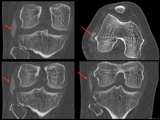
Traumatisme au Rugby il y a 5 jours.
Rupture proximale isolée du Ligament Collatéral Latréral (flèches blanches) avec persistanced'un moignon (petites flèches blanches) en regard du tubercule condylien latéral.
Suffusion hémoragique associée (*) des tissus sous cutanés en regard.
LCA normal (tête de flèche blanche).

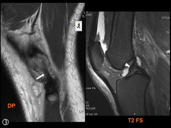
Homme de 45 ans présentant des douleurs médiales du genou droit post-traumatiques.
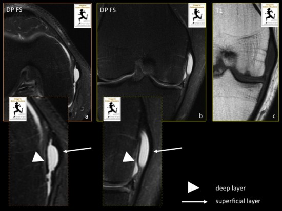
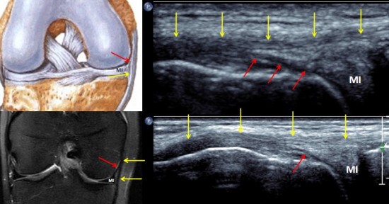
Les séquences IRM axiale et coronale DP avec saturation de la graisse (a,b) et coronale en pondération T1 (c) montrent une bursite sous la forme d’une collection liquidienne (hypersignal DP FS, Hyposignal T1) verticale située entre les portions profonde (faisceau ménisco-fémoral, tête de flèche) et superficielle (flèche) du ligament collatéral médial (LCM). On ne met pas en évidence de fissure méniscale en regard. On notera une ménisco-chondropathie fémoro-tibiale médiale associée.
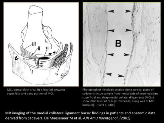
Causes des bursites du LCM: genu varum, arthrose avec ostéophytose médiale, polyarthrite rhumatoïde, pied plat, traumatisme et microtraumatismes.
MR imaging of the medial collateral ligament bursa: findings in patients and anatomic data derived from cadavers. De Maeseneer M et al. AJR Am J Roentgenol. (2001).
Anne Cotten, Imagerie musculosquelettique : Pathologies locorégionales, Masson.
Patient de 48 ans adressé pour gonalgie post-traumatique.
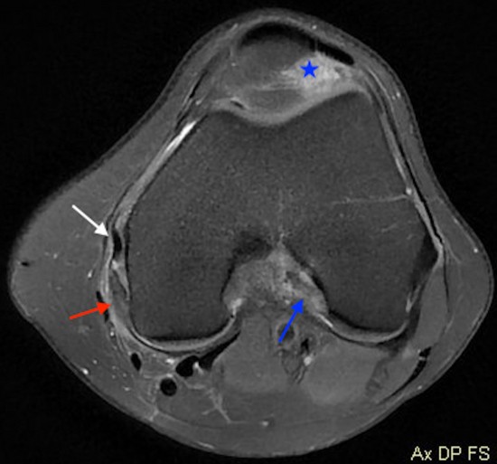
L'IRM en coupe axial DP FatSat retrouve une infiltration du plan ligamentaire médial prédominant en postérieur (, l'enthèse proximale du ligament oblique postérieur (flèche rouge) apparait épaissi en hypersignal T2, sans anomalie de l'enthèse proximale du ligament collatéral médial (flèche blanche).
Aspect oedemateux de l'insertion proximale du ligament croisé antérieur en rapport avec une rupture partielle (flèche bleue).
Infiltration de la graisse sous patellaire témoignant d'un conflit condylo-patellaire (étoile bleue).
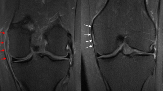
Les coupes coronales DP FatSat retrouvent l'aspect épaissi et en hypersignal du ligament oblique postérieur ( flèches rouges) sans avulsion de l'enthèse proximale, grade 2. Intégrité du ligament collatéral médial (flèches blanches).
Conclusion:
Lésion du plan ligamentaire médial avec atteinte isolée du ligament oblique postérieur, dans un contexte de rupture partielle du ligament croisé antérieur.
