Hanche | Pathologie labrale
L'arthro-IRM de hanche est l'examen de référence dans le diagnostic de lésion labrale du sujet jeune et présente des performances supérieures à celles de l'arthroscanner. Les lésions labrales sont souvent associées à un conflit fémoro-acétabulaire de type came chez le sujet jeune.
La topographie préférentielle est antéro-supérieure et les aspects sont variables:
- lésion du bord libre qui présente un aspect arrondi: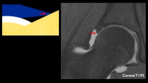
- fissuration +/-transfixiante visible grâce au passage de produit de contraste à partir de la face profonde
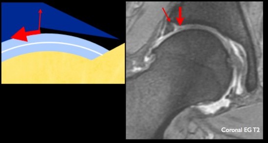
- lésion complexe (avec composantes verticale et horizontale) potentiellement instable
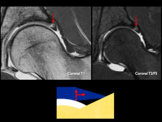
Dans le conflit fémoro-acétabulaire par effet came, ces anomalies s'accompagnent de lésions cartilagineuses de délimination c'est à dire développées à partir de la face profonde du cartilage acétabulaire (fléches épaisses). Ces lésions cartilagineuses apparaissent en hypersignal T2 en périphérie de l'os sous-chondral.
Références
Perpidakis E & al. Comparison of MR-arthrography and MDCT-arthrography of labral and articular cartilage pathology. Skeletal Radiol 2011; 40: 1441-1447.
Pfirrmann CW, Duc SR, Zanetti M, Dora C, Hodler J. MR arthrography of acetabular cartilage delamination in femoroacetabular cam impingement. Radiology. 2008 Oct;249(1):236-41.
Czerny & al. Lesions of acetabular labrum: accuracy of MR imaging and MR-arthrography in detection and staging. Radiology 1996; 200:225-30.
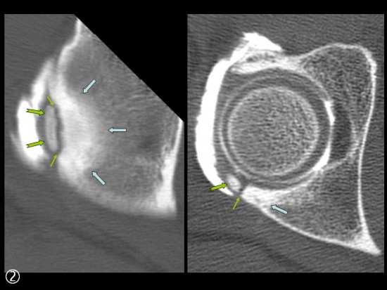
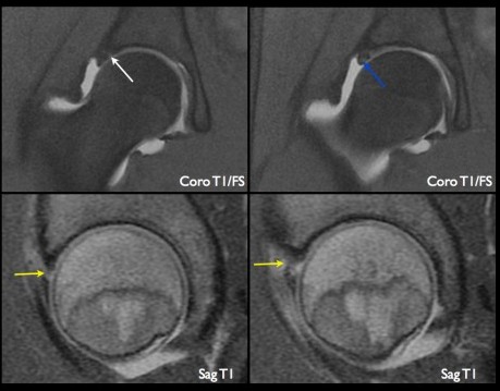
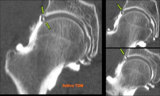
Patient de 26 ans, présentant un ressaut interne douloureux au niveau de la hanche droite.
Arthro-IRM:
- les coupes coronales objectivent une fissure supérieure (flèche blanche) ainsi qu'une atteinte du bord libre (flèche bleue).
- les coupes sagittales montrent une désinsertion proximale (flèches jaunes) du labrum cotyloïdien antérieur en rapport avec une lésion instable.
Douleur de hanche survenant essentiellement de Flexion-Abduction-Rotation Externe.
Fissuration du bourrelet cotyloïdien antérieur (![]() ) se poursuivant par un fin collet jusqu'a l'interligne articulaire (
) se poursuivant par un fin collet jusqu'a l'interligne articulaire (![]() ). Cartilages de revêtements sont réguliers (
). Cartilages de revêtements sont réguliers (![]() ).
).
Douleur intermittente avec sensation de ressaut de la hanche. IRM (hors centre) normale.
Fissuration longitudinale partiellement transfixiante (![]() ) du bourrelet cotyloïdien supérieur.
) du bourrelet cotyloïdien supérieur.
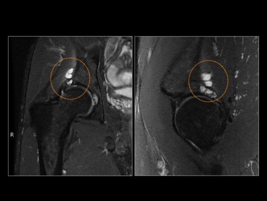
Douleur de hanche à l’abduction chez une femme de 56 ans présentant une dysplasie a minima .
Fissuration antéro supérieure (![]() ) d’un méga bourrelet cotyloïdien se prolongeant par un clivage (
) d’un méga bourrelet cotyloïdien se prolongeant par un clivage (![]() ). Géode sous chondrale du toit du cotyle opacifiée par le contraste (
). Géode sous chondrale du toit du cotyle opacifiée par le contraste (![]() ) associée à une chondropathie de stade III – IV de la tête fémorale (
) associée à une chondropathie de stade III – IV de la tête fémorale (![]() ).
).
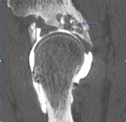
Fissure du labrum supérieur (flèche verte) de la hanche droite avec macrokyste (flèche bleue) en regard interessant le toit du cotyle.
Arthropathie dgénérative débutante avec chondropathie intéressant la tête fémorale et le toit du cotyle.
Pertite plage de délamination chondrale focalisée avec languette en avant de la fovéa (flèche rouge).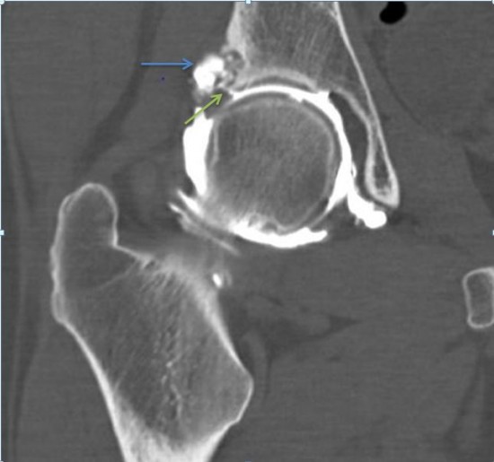
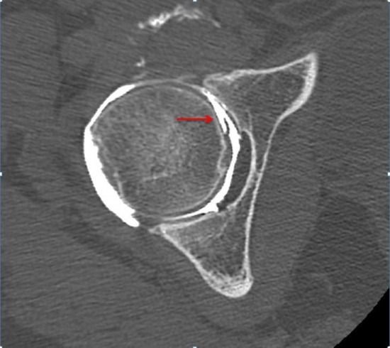
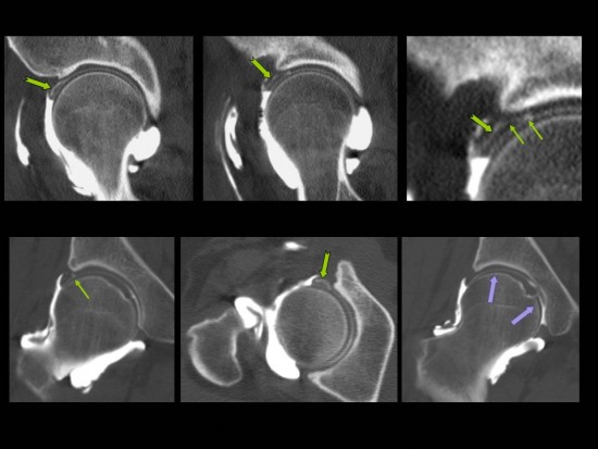
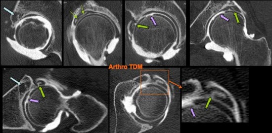
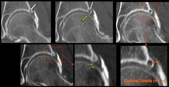
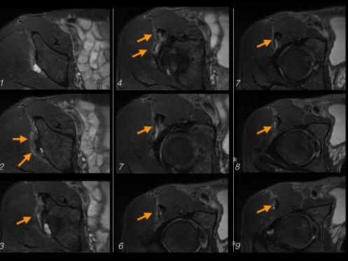
Douleur intermittente de l’aine chez un coureur de 400 m haies.
Mise en évidence sur l’arthroscanner d’une petite lacune de l’os sous chondral (![]() ) du toit du cotyle avec fine encoche chondrale en regard (
) du toit du cotyle avec fine encoche chondrale en regard (![]() ) et micro fissure du labrum (
) et micro fissure du labrum (![]() ). L’acquisition réalisée tardivement (+ 1 H) confirme la fissure avec présence de produit de contraste au sein de la lacune (
). L’acquisition réalisée tardivement (+ 1 H) confirme la fissure avec présence de produit de contraste au sein de la lacune (![]() ).
).
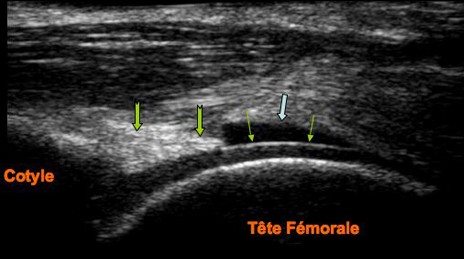
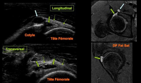
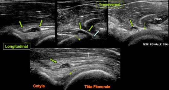
La présence d’un discret épanchement intra articulaire (![]() ) permet de souligner le cartilage de revêtement de la tête fémorale (
) permet de souligner le cartilage de revêtement de la tête fémorale (![]() ). Le bourrelet cotyloïdien se présente sous forme d’un triangle hyper échogène (
). Le bourrelet cotyloïdien se présente sous forme d’un triangle hyper échogène (![]() ) reposant sur le cotyle.
) reposant sur le cotyle.
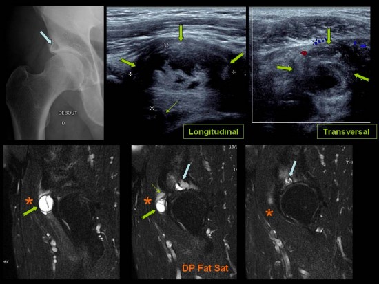
Douleur inguinale.
Volumineux kyste mucoïde hétérogène (![]() ) développé sous le muscle psoas (
) développé sous le muscle psoas (![]() ), en superficie d'un labrum lésé et remanié (
), en superficie d'un labrum lésé et remanié (![]() ), associé à des remaniements scléro kystiques de l'os sous chondral du cotyle (
), associé à des remaniements scléro kystiques de l'os sous chondral du cotyle (![]() ) et à un pincement de l'interligne articulaire.
) et à un pincement de l'interligne articulaire.
Douleur d’effort à l’aine chez une jeune sportive de 14 ans pratiquant l’athlétisme.
Structure kystique (![]() ) localisée au sein du bourrelet cotyloïdien antérieur (
) localisée au sein du bourrelet cotyloïdien antérieur (![]() ) se prolongeant en avant du pilier antérieur du cotyle (
) se prolongeant en avant du pilier antérieur du cotyle (![]() ). Absence d’anomalie intra articulaire à l’IRM.
). Absence d’anomalie intra articulaire à l’IRM.
Patient de 55 - sportif
Douleur racine de cuisse face antérieure d'évolution progressive
Demande d'échographie: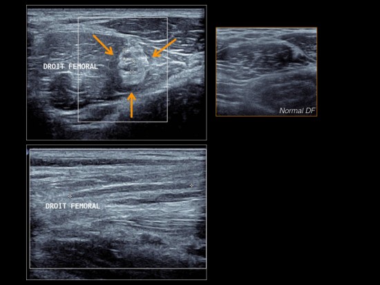
Sur cet examen on constate un net épaississement de la cloison centrale du muscle droit fémoral avec des remaniements microkystiques intralésionnels.
L'aspect fait évoquer en premier lieu des séquelles de lésion musculaire traumatique intrinsèque (de type désinsertion myo aponévritique central) avec cicatrice pathologique.
Une IRM est réalisée en complément.
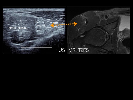
Sur ces images on voit la correspondance échographie / IRM et on confirme l'épaississement et les remaniements micro kystiques de la cloison centrale du droit fémoral.
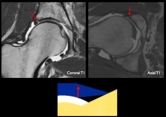
Les coupes coronales réalissées révèlent la présence d'un kyste d'origine labral en supéro externe. Il présente une composante intra osseuse et une composante au sein des parties molles adjacentes.
Les coupes axiales successives montrent que le kyste labral s'étend via le tendon indirect à la cloison central du muscle droit fémoral... l'image d'échographie est ainsi sans rapport avec une éventuelle séquelle de déchirure musculaire.
Douleurs de l’ aine survenant lors d’efforts sportifs.
Structure kystique (![]() ) se poursuivant par un collet (
) se poursuivant par un collet (![]() ) jusqu’au bourrelet cotyloïdien antérieur (
) jusqu’au bourrelet cotyloïdien antérieur (![]() ).
).
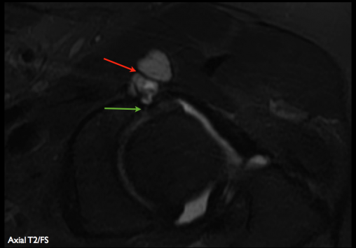
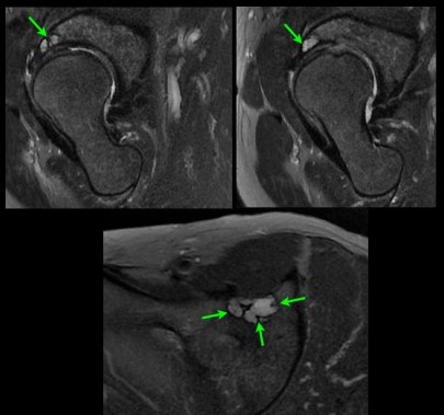
Patiente de 55 ans adressée pour douleurs inguinales de la hanche gauche et "image kystique" sur le bilan échographique. Arthro-IRM montrant la présence d'un kyste synovial d'aspect polylobé apparaissant en hypersignal T2 (flèches rouges) et développé à partir d'une fissure du du labrum cotyloïdien antérieur et supérieur (flèches vertes)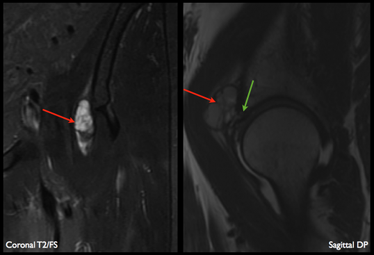
Patiente de 65 ans présentant une coxalgie chronique.
IRM: les séquences en pondération T2 montrent une formation kystique polylobée développée au niveau du toit du cotyle et à point de départ labral. Article T. Magee
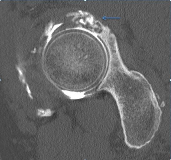
Sportif de 41 ans pratiquant le Jujitsu, se plaignant d'une douleur lors de l'élévation abduction rotation externe de la jambe. Pas de notion de traumatisme.
Le versant postérosupérieur du bourrelet cotyloïdien est le siège d'une ossification (![]() ) ou os acetabuli.
) ou os acetabuli.
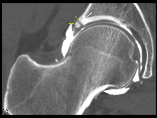
L'interligne avec le cotyle est discrètement hétérogène (![]() ) et le versant cotyloïdien présente une condensation (
) et le versant cotyloïdien présente une condensation (![]() ) en regard témoignant d'une instabilité à ce niveau.
) en regard témoignant d'une instabilité à ce niveau.