
Wrist | Arthritis | Calcium pyrophosphate dihydrate deposition disease of the wrist. X-Ray, CT, MRI
78-year-old patient, left wrist pain with seizures and pain free intervals.
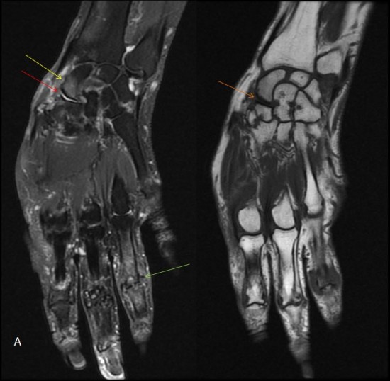
Figure A: MRI of the wrist, fat-sat DP coronal sequences and T1 coronal: scapho-trapezial arthropathies (red arrow) and interphalangeal proximal of the 4th finger (green arrow), congestive (bone edema, yellow arrow), and subchondral bone condesation (orange arrow).
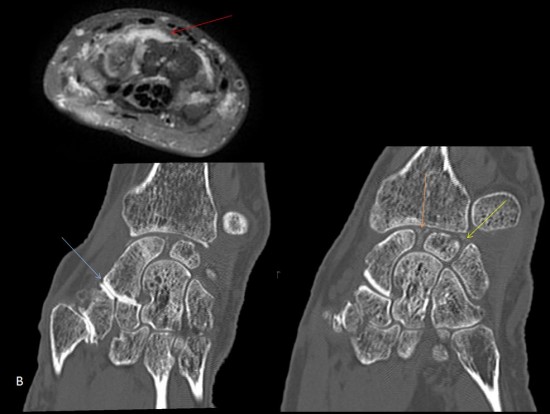
Figure B: MRI T1 Fat-Sat sequence with injection of gadolinium chelates and CT scan performed without injection: scapho-trapezial synovitis (red arrow), sub-chondral bone condesation (blue arrow), calcifications of the scapholunate and lunotriquetral ligaments (orange and yellow arrows, respectively).
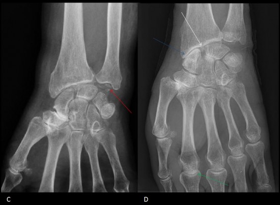
Figure C: Other patient: in addition to the signs already described, note the presence of chondrocalcinosis of the TFCC disk.
Figure D: Other patient: Pinching of the 2nd metacarpophalangeal (green arrow), and scapholunate diastasis (white arrow) testifying to a SCAC wrist stage II.
References:
-Steinbach LS. Calcium pyrophosphate dihydrate and calcium hydroxyapatite crystal deposition diseases: imaging perspectives. Radiol Clin North Am 2004 ; 42: 185-205.
- Belhouane R, Lebeau N, Maes-Clavier C, et al. Reproductibility of X-Rays and CT arthrography in SLAC, SNAC, SCAC wrists. Hand Surg Rehabil dec 2016 ; 35 (6) : 393-400.
