
Elbow | Instability | rupture of the ulnar collateral ligament : sonography and MRI
Traumatism of the left elbow in a 32 year old man.


Figure 1 and 2: normal longitudinal sonography of common flexor tendon (blue arrow) and ulnar collateral ligament (green arrow)

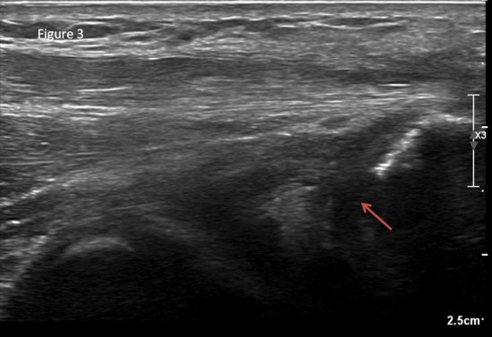
Figure 3 : longitudinal sonography: proximal rupture of the ulnar collateral ligament (red arrow)
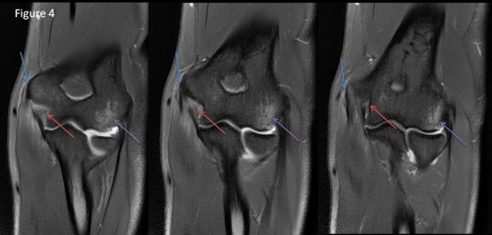
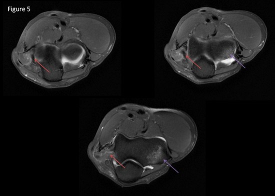
Coronal (figure 4) and axial (figure 5) fat suprression MRI
- proximal rupture of the ulnar collateral ligament (red arrow)
- contusion of lateral condyle of humérus (violet arrow)
- normal common flexor tendon (blue arrow)
Miller TT, Alder RS, Friedman L. Sonography of injury of the ulnar collateral ligament of the elbow-initial experience. Skeletal Radiol 2004
Brunton LM, Anderson MW, Pannunzio ME, Khanna AJ, Chhabra AB. Magnetic resonance imaging of the elbow : uptade on current techniques and indications. J Hang Surg Am. 2006; 31:1001-1011
