
Shoulder | Arthritis | Primary synovial chondromatosis: US, MRI and CT
Pain of the right shoulder in a young male of 31 years old
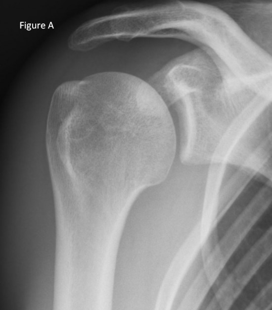 Figure A: normal x-ray of the right side shoulder
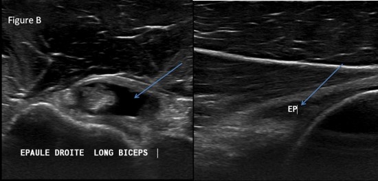 Figure B: ultrasound of right shoulder
Intra-articular effusion (blue arrow) visualized within the biciipital groove and within the posterior articular recess
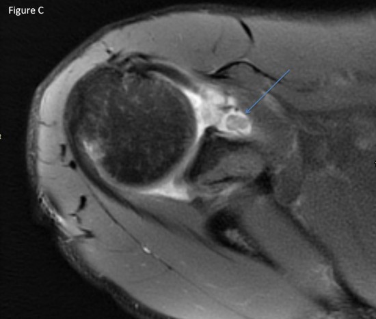
Figure C: axial MRI DP fat SAT
Significant intra-articular effusion communicating with the bicipital gutter, of heterogeneous appearance, which may be compatible with the presence of osteochondromas (blue arrow).
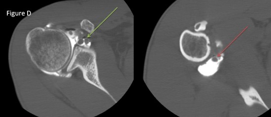
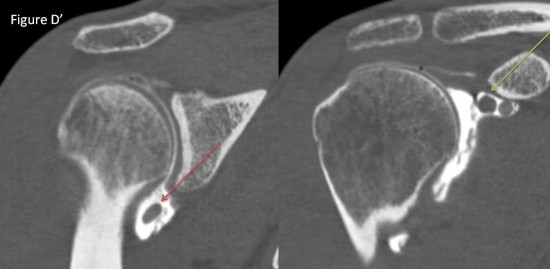
Figure D et D': axial (D) and frontal (D') CT
Non-ossified and non calcified multiple rounded formations in the lower joint recess (red arrow) and the sub-coracoid recess (green aroow), suggesting primitive osteochondromatis.
