
Knee | Cruciate ligaments | Anteromedial Meniscofemoral Ligament (AMMF) : MRI
50 year old patient presenting with left knee pain : Fortuitous discovery of an AMMF.

Images (a), (b),(c) et (d) : DP FS weighted sagittal views of the left knee showing an AMMF (blue arrows), situated in front of the ACL (red arrow), inserting on the anterior aspect of the medial meniscus (black arrow), close to the insertion of the menisco meniscal ligament (green arrow). Orange arrow : PCL.
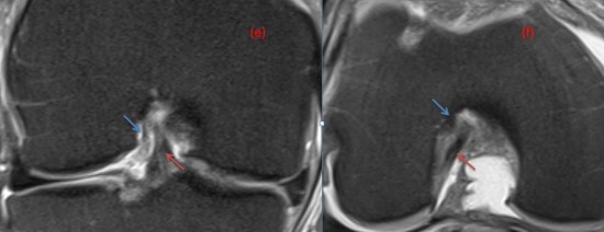
Image (e) DP FS weighted coronal view, showing the AMMF (blue arrow) situated laterrally to the ACL (red arrow).
Image (f) DP FS weighted axial view, showing the AMMF (blue arrow) situated laterrally and anteriorly to the ACL (red arrow) on the intercondylar fossa.
This case illustrates an anteromedial meniscofemoral ligament, a rare variant whose prevalence is estimated at <1%.
Arthroscopy. 2018 May;34(5):1590-1600. doi: 10.1016/j.arthro.2017.12.010.
