
Foot | Arthritis | Freiberg's disease
14 year-old female patient, presenting with chronic foot pain, with normal initial plain radiographs. MRI exploration :
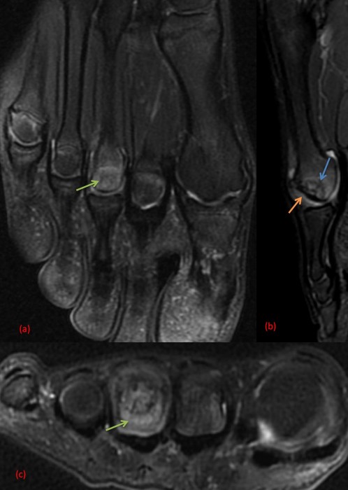
T2 FAT SAT weighted slices of the front foot : (a) axial slice showing edema of the 3rd metatarsal head (green arrow), (b) sagittal slice showing a serpigineous low intensity signal going from cortical to cortical (blue arrow) associated with sub chondral sinking of the 3rd metatarsal head (orange arrow), (c) corresponding axial slice
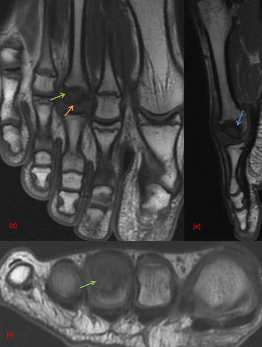
T1 weighted corresponding slices : (d) axial slice : edema (green arrow) and sub chondral sinking with slight deformation of the 3rd metatarsal head (orange arrow), (e) sagittal slice : sinking of the metatarsal head with serpigneous line going from cortical to cortical (blue arrow), (f) corresponding axial slice
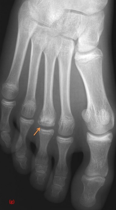
Investigation has been completed with a plain radiograph, confirming the subchondral sinking of the 3rd metatarsal head (orange arrow). No joint space narrowing.
This case illustrates Freiberg's disease (Kohler's second disease), which corresponds to an aseptic osteonecrosis, involving most frequently the 2nd metatarsus but also the 3rd one.
This disease affects frequently the young female population, at the adolescence.
Etiology is poorly understood, with the hypothesis of an avascular necrosis favoured by microtraumatic factors and foot disbalance (too long metatarsus).
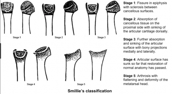
Smillie has proposed a classification for this pathology :
In the early stage, a simple orthopedic treatment may be followed but in case of failure to heal or evolved stage disease, a dorsiflexion osteotomy is indicated.
Smillie I (1957) Freiberg’s infraction (Kohler’s second disease).J Bone Joint Surg 39B:580
https://www.orthobullets.com/foot-and-ankle/7018/freibergs-disease
